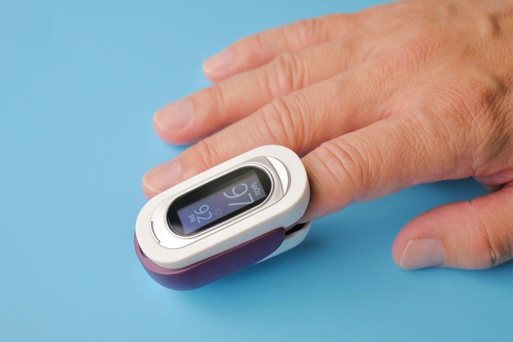
SpO2 jumping from 98% to 83% is alarming.
A fingertip oximeter that flips between 98%, 86% and 99% triggers panic, doubt about the device, and a decision: ignore, retest, or seek care. Those reactions are normal.
Most sudden dips are noise — motion, poor perfusion, cold fingers, nail polish. Use a simple rule: brief dips (<15 sec) with no symptoms and a steady pulse likely are artifacts; persistent lows (>60 sec), mismatched pulse, or symptoms require escalation.
Retest twice after warming and repositioning.
Compare oximeter pulse to radial pulse.
Remove polish; avoid movement.
Seek help if SpO2 <90% persistently or symptoms.
How transmissive/reflective oximetry and the pleth produce SpO2 and pulse rate
- Transmissive vs reflective sensors
Transmissive sensors pass red and infrared light through thin tissue (finger or earlobe); reflective sensors measure back‑scattered light from surfaces (wrist, forehead). Both use two LEDs and a photodiode to capture wavelength‑dependent absorption.
- Two wavelengths and the measurement principle
Oxygenated and deoxygenated haemoglobin absorb red and infrared light differently; the device isolates the pulsatile (AC) absorption from the steady (DC) component and computes a ‘ratio of ratios’ that maps, via calibration, to SpO2.
- The plethysmograph signal
The photodiode trace (pleth) contains a DC baseline plus an AC pulsatile waveform from arterial blood volume changes; AC frequency gives pulse rate and AC amplitude reflects pulse strength.
- Why a clean pulsatile signal matters
SpO2 is derived from the AC amplitudes at both wavelengths, so a clear, regular pleth is essential for accuracy; motion, low perfusion, venous pulsation, ambient light or poor contact reduce AC amplitude or add noise, causing fluctuating readings.
- Practical markers of trustworthy readings
Reliable readings show regular pleth peaks matching the displayed pulse rate and a stable baseline; a small, noisy or irregular pleth suggests the SpO2 value is likely artifact until corroborated.
Low‑perfusion pattern
Recognizing low perfusion
A weak or absent pleth (flat, noisy, or barely pulsatile waveform) often produces a drifting or persistently low SpO2 reading while the user remains asymptomatic. At the finger this appears as a cold, pale or clammy digit, faint pulse on palpation, and SpO2 that wanders independent of breathing.
Practical, reversible fixes
Try these steps in sequence and re-check the pleth after each:
- Warm the hand: rub, soak briefly in warm water, or place the arm below heart level to increase flow.
- Change or reposition the probe: switch fingers, ensure correct alignment, avoid tight straps.
- Remove barriers: take off nail polish, artificial nails, rings; clean the sensor and finger.
- Reduce motion and ambient light: sit still and shield the sensor from direct bright light.
- Stimulate perfusion: gentle massage or squeezing the finger, or brief light activity to raise cardiac output.
If readings improve after these maneuvers, low perfusion was likely the cause; persistent poor pleth needs clinical evaluation.
If the pleth is flat but the person feels well, warm and reposition the finger first. A quick finger swap and rubbing usually restores a usable waveform.
Motion and external artifact
Motion produces a distinctive signature: SpO2 and pulse rate often jump together while the pleth waveform becomes irregular — baseline wander, jagged peaks, or dropped pulses. Transient drops that coincide with visible movement are usually artifact rather than true desaturation.
How to confirm motion artifact
Inspect the pleth and pulse rate simultaneously. A reliable pleth shows regular, evenly spaced pulsatile peaks; motion makes that pattern noisy or intermittent. If SpO2 spikes or dips align with a chaotic pleth and a sudden pulse-rate change, motion artifact is the most likely cause.
Practical stabilisation techniques
- Rest and support the limb; eliminate swinging or muscle tension.
- Use light adhesive or medical tape to secure the probe and cable.
- Try a different finger or a forehead/reflectance probe for restless subjects.
- Warm cold fingers, remove nail polish, and reduce bright ambient light.
- For overnight recording, correlate spikes with movement using an accelerometer or consider home sleep-monitoring devices that log motion.
If disturbances are brief and pleth-disordered, interpret spikes as artifact; persistent low SpO2 with a stable pleth needs clinical escalation.
Optical and environmental causes of fluctuation
Several non‑physiological factors frequently produce inconsistent SpO2 readings. Nail polish and artificial nails alter light transmission; strong ambient light can flood the photodiode. Sensor site issues (poor perfusion, thick tissue) degrade the pulsatile signal, and skin pigmentation can change how sensors calibrated on lighter skin perform.
Quick tests
- Remove polish or try a different finger — a rapid change implicates nail or site.
- Cover the sensor with a hand or cloth to block external light; if the signal steadies, ambient light was the problem.
- Compare the oximeter pulse rate to a reliable pulse source; mismatches suggest motion or weak pleth.
Low‑effort fixes
- Remove nail barriers, warm and massage the finger, or switch to an alternate site (ear/forehead).
- Shield the probe from direct light or secure it with tape/cloth.
- For persistent issues, choose devices validated for diverse skin tones or consult clinical accuracy studies and regulator guidance for deeper evaluation.
Device, placement, and maintenance checks
If SpO2 or pulse fluctuates while symptoms and clinical context are stable, the unit or its use is often to blame. Common signs of device fault include a flat or noisy pleth waveform, readings that jump with slight finger movement, pulse rate discordant with a known heart rate, or intermittent power/battery issues.
Quick troubleshooting steps:
- Inspect and clean contact surfaces; remove nail polish or thick acrylics.
- Replace or recharge batteries; reseat any detachable sensor.
- Try a different finger or ear lobe and ensure the probe fits snugly.
- Restart the unit and observe the pleth waveform for stable pulsatility.
- Compare with a second oximeter or clinical monitor to confirm disagreement.
For buying guidance, prefer devices with visible plethysmogram, motion‑tolerance features, replaceable probes and regulatory clearance; see the broader list of home health monitors you need for recommendations.
True physiologic fluctuation vs artifact
Brief, transient SpO2 dips are usually artifact; true physiologic fluctuation is sustained, reproducible, or accompanied by other signs. Use the pleth and context to decide.
Objective cues that indicate true physiologic change
- Sustained desaturation (SpO2 below expected baseline for >60–120 seconds) with a stable pleth waveform.
- Concurrent physiologic signs: new or worsening dyspnea, tachypnea, chest pain, altered mental status, cyanosis, or hemodynamic instability.
- Concordant vital sign changes: oxygen saturation fall accompanied by pulse‑rate changes on the monitor and clinical correlation.
- Reproducibility across sites/devices: low readings on alternate fingers or a second oximeter.
Short escalation algorithm
- Rapid troubleshooting: confirm pleth quality, reposition or change probe, warm site, remove nail barriers, and retest another finger or forehead.
- If low values persist or symptoms/signs are present, treat per local protocol (oxygen, airway support) and escalate to urgent clinical evaluation or emergency services.
Any of the following warrants urgent action:
SpO2 ≤ 90% sustained despite troubleshooting New respiratory distress, chest pain, syncope, or altered consciousness Rapid desaturation with hemodynamic compromiseIf present, treat and arrange emergency assessment without delay.
Step-by-step troubleshooting checklist
-
Inspect pleth and symptoms
Confirm dips coincide with pleth distortion or symptoms; note time, duration, and whether pulses on the plethogram remain regular.
-
Stabilize the sensor site
Warm the finger, remove polish or artificial nails, clean skin, and reposition the probe for a snug, even contact.
-
Minimize motion and light
Support the hand, secure the probe with tape or strap, and shield from bright ambient light or camera flashes.
-
Compare sites and devices
Swap fingers, test earlobe or forehead, and compare readings with a second validated oximeter or pulse monitor.
-
Power-cycle and inspect hardware
Restart the unit, replace batteries, check cables and contacts for wear or loose parts.
-
Document and escalate
Record pleth traces during tests; if artifacts persist at rest across sites, prepare to replace or service the device.
How to reproduce faults reliably
Reproduce both a controlled rest and deliberate motion test. Start with a 2‑minute baseline while seated and breathing normally, recording pleth stability and SpO2 variance. Then perform standardized motions: slow finger wiggle, firm taps every 5–10 seconds, and a 30‑second walk. Repeat tests on an alternate site (earlobe or forehead).
Consistent artifacts during rest indicate contact or device fault; artifacts only during motion point to motion sensitivity.
When to replace the device
Replace when any apply:
- Persistent unexplained SpO2 drops at rest despite good perfusion.
- Absent or flat pleth while the finger is warm and still.
- Frequent failures across multiple sites or comparison devices.
- Visible damage, loose optics, or unstable power/battery behavior.
Consider replacement for clinical reliability if the unit is older than 2–3 years or firmware/service cannot resolve faults.
How long to wait between re-tests?
Wait 30–60 seconds after movement to let the pleth stabilise. If the trace remains unstable, warm the extremity or change the probe site and retest.
When is a second oximeter worth buying?
Purchase a second medically cleared oximeter when checks (different finger, clean sensor, restart) still show inconsistent pleth or persistent disagreement. Comparing two units helps determine device fault versus true physiology.
How many retries before escalation?
Three reproducible low readings or any symptomatic desaturation warrants escalation. Record the waveform, site, and interventions when seeking care.
Can smartphone or cheap probes be trusted?
Smartphone or cheap probes can indicate trends but often lack consistent accuracy. Confirm critical or unexpected readings with a clinical-grade device.
FAQ and Practical Rule
- Allow 30–60 s for the pleth to settle before trusting a re-test
- Use waveform review and a second validated device to confirm suspect readings
- Document and escalate when lows are reproducible or accompanied by symptoms
Rule: reproduce, stabilise, escalate if symptomatic. Reproduce the event deliberately, stabilise the signal (warm, reposition, remove artifacts), and escalate care for persistent drops or any associated symptoms.